HUMAN ANATOMY
PELVIS AND PERINEUM
(Lecture Notes)
(Lecture Notes)
Necdet Ersöz
Gazi University Medical School
Gazi University Medical School
Bony Pelvis
Composition: formed by paired hip bones, sacrum, coccyx, and their
articulations.
Two portions: Greater and Lesser pelvis
Lesser pelvis:
ü
Terminal
line (pelvic inlet): formed by promontory of sacrum, arcuate line, pectin of
pubis, pubic tubercle, upper border of pubic symphysis.
ü
Pelvic
outlet: formed by tip of coccyx, sacrotuberous ligament, ischial tuberosity,
ramus of ischium, inferior ramus of pubic symphysis.
Muscles of Pelvic Wall: Piriform muscle and Obturator internus muscle
Anteroinferior pelvic wall is formed primarily by the bodies and rami of the
pubic bones and the pubic symphysis. It participates in bearing the weight of
the urinary bladder.
Lateral pelvic wall is formed by the right
and left hip bones -includes an obturator foramen caused by an obturator
membrane >> obturator internus
muscles cover and thus pad most of the lateral pelvic walls. The fibers of
each obturator internus converge posteriorly, become tendinous, and turn
sharply laterally to pass from the lesser pelvis through the lesser sciatic foramen to attach to the greater trochanter of the femur. The medial surface of these
muscles is covered by obturator fascia,
thickened centrally as a tendinous arch that provides attachment for the pelvic
diaphragm.
Posterior pelvic wall is, in the anatomical position, consists of a bony
wall and roof in the midline (formed by the sacrum and coccyx) and musculoligamentous posterolateral walls,
formed by the ligaments associated with the sacroiliac joints and piriformis muscles.
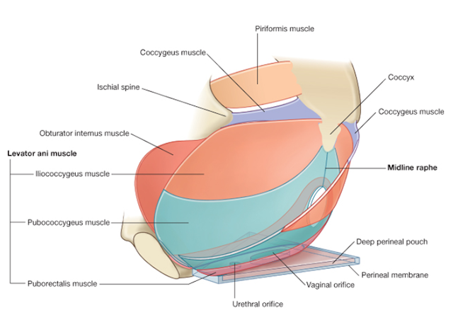
Pelvic Floor
ü
Formed
by the bowl – or – funnel-shaped pelvic
diaphragm, which consists of the coccygeus and levator ani muscles and the
fascias covering the superior and inferior aspects of these muscles.
ü
The
pelvic diaphragm lies within the lesser pelvis, separating the pelvic cavity from the perineum, for which it forms
the roof .
ü
The
attachment of the diaphragm to the obturator fascia divides the obturator
internus into a superior pelvic portion and an inferior perineal portion.
Pelvis: superior fascia of pelvic diaphragm, levator ani
muscle, coccygeus, and inferior fascia of pelvic diaphragm.
Muscles of floor of pelvis
and pelvic diaphragm
ü
Lavator
ani muscle: puborectalis, pubococcygeus, and iliococcygeus
ü
Coccygeus
Coccygeus
ü Arise from the lateral
aspects of the interior sacrum and coccyx, their fleshy fibers lying on and
attaching to deep surface of the
sacrospinous ligament.
Levator ani muscle
(puborectalis, pubococcygeus, and iliococcygeus)
ü Proximal
attachment: body of
pubis, tendinous arch of obturator fascia, ischial spine
ü Distal
attachment: perineal
body, coccyx, anococcygeal ligament, walls of prostate or vagina, rectum, and
anal canal.
ü Forms most of pelvic diaphragm that helps support
pelvic viscera and resists increases in intra-abdominal pressure.
ü The levator ani muscles help support the pelvic viscera and maintain
closure of the rectum and vagina. They are innervated directly by branches
from the anterior ramus of S4 and by branches of the pudendal nerve (S2 to S4)
ü
An
anterior gap between the medial borders of the levator ani muscles of each side
– the urogenital hiatus – gives passage to the urethra and, in females, vagina.
Puborectalis:
ü
The
thicker, narrower, medial part of the levator ani muscle.
ü
It
forms U-shaped muscular sling
(puborectal sling) that passes posterior to the anorectal junction,
bounding the urogenital hiatus.
ü
This
part plays a major role in maintaining fecal continence.
Pubococcygeus:
ü
The
wider but thinner intermediate part of the levator ani, which arises from lateral
to the puborectalis.
ü
Its
lateral fibers attach to the coccyx and its medial fibers merge with those of
the contralateral muscle to form a fibrous
raphe or tendinous plate, part of the anococcygeal body or ligament between
the anus and the coccyx (often referred to clinically as the “levator plate”)
ü
Shorter
muscular slips of the pubococcygeus extending medially and blending with the
fascia around midline structures are named for the structure near their termination:
pubovaginalis (females), puboprostaticus (levator prostate, males),
puboperinealis, puboanalis.
Iliococcygeus:
ü
The
posterolateral part of the levator ani, which arises from the posterior tendinous arch and ischial spine.
ü
It
is thin and often poorly developed (appearing more aponeurotic than muscular)
and also blends with the anococcygeal body posteriorly.
Pelvic Fascia (Parietal
pelvic fascia + Visceral pelvic fascia)
Parietal pelvic fascia:
ü
A continuation of the transverse fascia into the
pelvis. It covers the piriformis and obturator internus
muscle.
ü
Attaches
to the arcuate line of the pubis and
ilium, thickens over the obturator internus muscle to form the arcus tendinous, the origin of portions
of the levator ani muscle.
ü
At
the tendinous arch of levator ani it splits to cove both superior and inferior surfaces
of the levator ani as superior and inferior fascia of pelvic diaphragm.
Visceral pelvic fascia:
ü
Lies
between the peritoneum and the pelvic viscera.
ü
It
is a continuation of the extraperitoneal connective tissue.
ü
Ensheathes
retroperitoneal viscera and forms septa between retroperitoneal organs:
rectovesical septum, rectovaginal septum
Retropubic space:
ü
Lies
between pubic symphysis and urinary bladder.
Pararectal space:
ü
Lies
around the rectus
ü
Retrorectal
space
Perineum
General Features
ü
Region
of below pelvic diaphragm
ü
A
diamond shape space whose boundaries are those of the pelvic outlet: lower border of symphysis pubis, rami of
pubis and ischium, ischial tuberosities, sacrotuberous ligament, the coccyx
Superficial Perineal Space
ü
A
potential space between the perineal fascia and the perineal membrane, bounded
laterally by the ischiopubic rami.
Boundaries of superficial
perineal space:
ü
Lies
between inferior fascia of urogenital diaphragm and superficial fascia of
perineum.
ü
Space
open anteriorly (in rupture of cavernous part of urethra, urine can extravasate
from scrotum upward in front of symphysis pubis into anterior abdominal wall
deep to membranous fascia of Scarpa)
Contents
ü
Posterior
part: superficial transverse perineal muscle
ü
Lateral
part: crus penis (male), crus of clitoris (female) and ischiocavernosus
covering them.
ü
Central
part: bulb of urethra (male), bulb of vestibule (female) and bulbocavernosus
covering them.
ü
Branches
of pudendal nerves and internal pudendal vessels.
In males, the superficial
perineal pouch contains the:
ü
Root
(bulb and crura) of the penis and associated muscles (ischiocavernosus and bulbospongiosus)
ü
Proximal
(bulbous) part of the spongy uretra
ü
Superficial
transverse perineal muscles
ü
Deep
perineal branches of the internal pudendal nerves
In females, the superficial
perineal pouch contains the:
ü
Clitoris
and associated muscles (ischiocavernosus)
ü
Bulbs
of the vestibule and surrounding muscle (bulbospongiosus)
ü
Greater
vestibular glands
ü
Superficial
transverse perineal muscles
ü
Related
vessels and nerves (deep perineal branches of the internal pudendal vessels
pudendal vessels and nerves)
Deep Perineal Space
ü
Lies
between superior and inferior fascia of urogenital diaphragm.
Contents
ü
Deep
transverse perineal muscle
ü
Bulbourethral
gland (male)
ü
Sphincter
of urethra (male), urethrovaginal sphincter
ü
Arteries,
veins and nerves
The perineal membrane
ü
The
perineal membrane is a thick fascial, triangular structure attached to the bony
framework of the pubic arch
ü
It
is oriented in the horizontal plane and has a free posterior margin.
ü
Anteriorly
there is a small gap between the membrane and the inferior pubic ligament (a
ligament associated with the pubic symphysis)
ü
The
perineal membrane is related above to a thin space called the deep perineal pouch (deep perineal space), which contains a
layer of skeletal muscle and various neurovascular elements.
ü
The
deep perineal pouch is open above and is not separated from more superior
structures by a distinct layer of fascia.
ü
The
parts of perineal membrane and structures in the deep perineal pouch, enclosed
by the urogenital hiatus above, therefore contribute to the pelvic floor and
support elements of the urogenital system in the pelvic cavity.
ü
The
perineal membrane and adjacent pubic arch provide attachment for the roots of
the external genitalia and the muscles associated with them.
ü
The
urethra penetrates vertically through a circular hiatus in the perineal
membrane as it passes from the pelvic cavity, above, to the perineum, below.
ü
In
women, the vagina also passes through a hiatus in the perineal membrane just
posterior to the urethral hiatus.
ü
In
both women and men, a deep transverse perineal muscle on each side parallels
the free margin of the perineal membrane
and joins with its partner at the midline.
ü
These
muscles are thought to stabilize the
position of the perineal body, which is a midline structure along the
posterior edge of the perineal membrane.
Perineal Central Tendon
ü
Wedge-shaped
fibromuscular mass
ü
In
female, between anal canal and lower end of vagina
ü
In
male, between anal canal and root of penis
ü
It
is larger in female than in male and five support to the posterior wall of the
vagina
Perineal Body
ü
An
ill-defined but important connective tissue structure into which muscles of the
pelvic floor and the perineum attach.
ü
It
is positioned in the midline along the posterior border of the perineal
membrane, to which it attaches.
ü
Origin
or insertion of several small muscles and insertion of part of pelvic diaphragm
ü
These
muscles are: sphincter ani externus, levator ani, superficial transverse muscle
perineum, deep transverse muscles perineum, bulbocavernousus, sphincter of
urethra (male) or urethrovaginal sphincter (female).
In both sexes, the deep
perineal pouch contains:
ü
Part
of the urethra, centrally.
ü
The
inferior part of the external urethral sphincter muscle, above the center of
the perineal membrane, surrounding the urethra.
ü
Anterior
extensions of the ischioanal fat pads.
In males, the deep perineal
pouch contains:
ü
Intermediate
part of the urethra, the narrowest part of the male urethra.
ü
Deep
transverse perineal muscles, immediately superior to the perineal membrane (on
its superior surface), running transversely along its posterior aspect.
ü
Bulbouretral
glands, embedded within the deep perineal musculature.
ü
Dorsal
neurovascular structures of the penis.
In females, the deep
perineal pouch contains:
ü
Proximal
part of the urethra.
ü
A
mass of smooth muscle in the place of deep transverse perineal muscles on the
posterior edge of the perineal membrane, associated with the perineal body.
ü
Dorsal
neurovasculature of the clitoris
Two Triangles
ü
An
imaginary line drawn between the two ischial tuberosities divides perineum into
anterior and posterior triangles.
ü
Urogenital
region (anterior) differs in male and female.
ü
Anal
region (posterior) similar in both sexes.
Anal Region
ü
Internal Anal Sphincter: smooth muscle (thickened circular muscle coat),
surrounds upper two-thirds of anal canal, autonomic nerve supply.
ü
External Anal Sphincter: striated muscle, surrounds lower two-thirds of anal
canal, three parts (subcutaneous, superficial, and deep), innervation by anal
nerves of pudendal nerves and branches of S4.
Ischioanal Fossa
ü
Paired,
wedge-shaped, fat-filled spaces on either side of anal canal.
Boundaries:
ü
Apex
– conjunctive area of inferior fascia of pelvic diaphragm and fascia covering
the obturator internus.
ü
Base
– skin of anal region.
ü
Medial
– sphincter ani externus, levator ani, coccygeus and inferior fascia of pelvic
diaphragm
ü
Lateral
– ischial tuberosity, obturator internus and fascia
ü
Anterior
– posterior border of urogenital diaphragm. Forward projection of anterior
recess of fossa between pelvic diaphragm above and urogenital diaphragm below.
ü
Posterior
– backward projection of posterior recess of fossa between gluteus maximus,
sacrotuberous ligament and coccyx.
Contents
ü
Fat
ü
Internal
pudendal artery and vein and their rectal branches.
ü
Pudendal
nerve and its inferior rectal branch.
Vessels and nerves enter from gluteal region, through
lesser sciatic foramen, travel on a fascial canal – the pudendal canal
(Alcocks) – on the lateral wall of fossa, and extend forward into urogenital
region.
Urogenital Region
Superficial fascia has two
layers:
ü
The
superficial or fatty layer
ü
The
deep or membranous layer (superficial fascia of perineum or Colles fascia)
ü
Anteriorly
it is continuous with Dartos of the
scrotum, fascia of the penis, membranous layer of superficial fascia of the
abdominal wall known as the fascia of Scarpa.
Deep fascia has two layers:
ü
Superior
fascia of urogenital diaphragm.
ü
Inferior
fascia of urogenital diaphragm.
Urogenital Diaphragm
ü
Triangular
in shape
ü
Attached
laterally to ischiopubic rami and ischial tuberosities.
ü
Formed
by sphincter of urethra, deep transverse perineal muscle, superior and inferior
fascia of urogenital diaphragm.
Note: These notes are taken from
Gazi University Faculty of Medicine Prof. Dr. Meltem BAHCELIOGLU's Anatomy lectures.


Yorumlar
Yorum Gönder
Görüş, öneri, soru ve eleştirilerinizi lütfen bildiriniz. Yapıcı yorumlar değerlendirilecek; kişilik saldırıları ve üslûp hataları engellenecektir.